CT Enterography – Procedure, Uses And Risks
Your healthcare provider can use CT enterography to determine what is causing your condition.
Author:Suleman ShahReviewer:Han JuJul 01, 202241 Shares1.1K Views

CT enterographyproduces images of the small and large bowels or colon. CT enterography can help doctors diagnose inflammatory bowel disease, gastrointestinal bleeding, and some gastrointestinal tumors. Fluid is taken that dilates the small bowel as part of the CT enterography exam. During the scan, an intravenous contrast agent makes blood vessels visible on the images and identifies bowel inflammation. A glucagon IV is used to slow bowel movement. To improve bowel filling, Reglan is taken orally.
What Is CT Enterography?
Enterography is derived from the terms"entero" (intestine or gut) and "graphy" (picture). CT enterography is an imaging technique that examines the small intestine using CT pictures and a contrast substance. The method enables your doctor to establish what is causing your problem. He or she can also tell you how well you respond to therapyfor a medical condition like Crohn's disease.
CT enterography is a rapid, painless, and precise treatment.
Unlike traditional X-ray pictures, CT enterography may offer detailed views of tissue and structures such as bone and blood arteries.
CT Enterography Indications
Among the indications for CT enterography are:
- Diagnosis and consequences of Crohn's disease (primarily)
- The most frequent reason is suspected small intestinal hemorrhage, generally conducted after a negative endoscopy for a suspected small intestine tumor, such as carcinoid or polyposis syndromes.
- Examine for problems such as lymphoma-related partial minor intestinal blockages, such as surgical adhesions, radiotherapy enteritis, and scleroderma.
- Persistent mesenteric ischemia is suspected
- Chronic diarrhea and stomach discomfort.
What Is Enterography?
CT Enterography Procedure
During CT enterography, images of cross-sections or slices of your abdominal tissues, focused on the small intestine, are obtained. Before the scan, you will consume a special kind of weak barium and get an injection in a vein through an IV to highlight the small intestine even more. CT scans may aid in the early detection of a diagnosis. Your doctor will use this information to identify the best therapy for you.
Get Ready
- Eat or drink nothing for 4 hours before this test.
- You will be outfitted with a gown and scrub trousers.
- For one week before the CT scan, no barium investigations should be performed.
- If you have a historyof sensitivities to contrast, see your doctor before the test.
- If you have not had a recent creatinine blood test, a finger-poke blood test may be performed before your scan.
During The Test
CT Enterography with contrast takes around 1 hour 45 minutes. This includes 45 minutes for the test and 1 hour for the barium to be consumed. Before you begin, the radiology technician will describe the CT scan in contrast to you. If you don't comprehend anything, ask questions. Before the examination, remove all clothes and change into a hospital gown and slacks. The technician will start an IV. The contrast material will be administered by IV. Contrast material will be administered via the IV.
The CT scan is not harmful. When the IV is begun, you will feel a prick. As the contrast material is administered, many individuals experience a warm "flush." This is common and will pass fast.
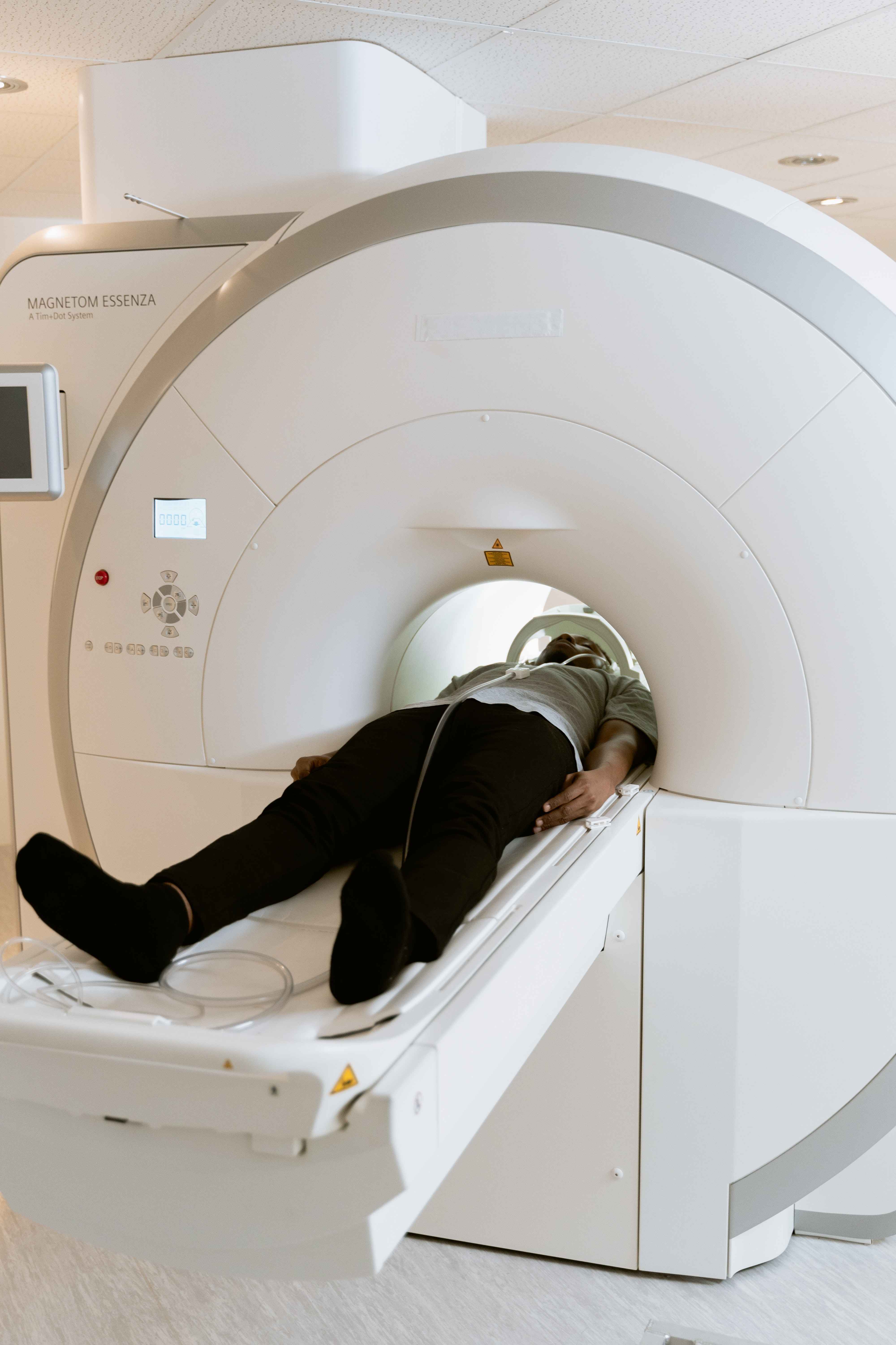
The CT scanner has a table on which you will lay and a doughnut-shaped ring. You will remain still on the table while it moves through the ring. During the exam, the technician will give you instructions. You will sometimes be required to lift your arms over your head. In addition, you will be asked to hold your breath for 10 to 12 seconds. While you hold your breath, the table will travel around the ring, taking images. The photographs will be taken before, during, and after the injection of contrast material into the IV. It is critical to remain motionless while the photographs are being shot.
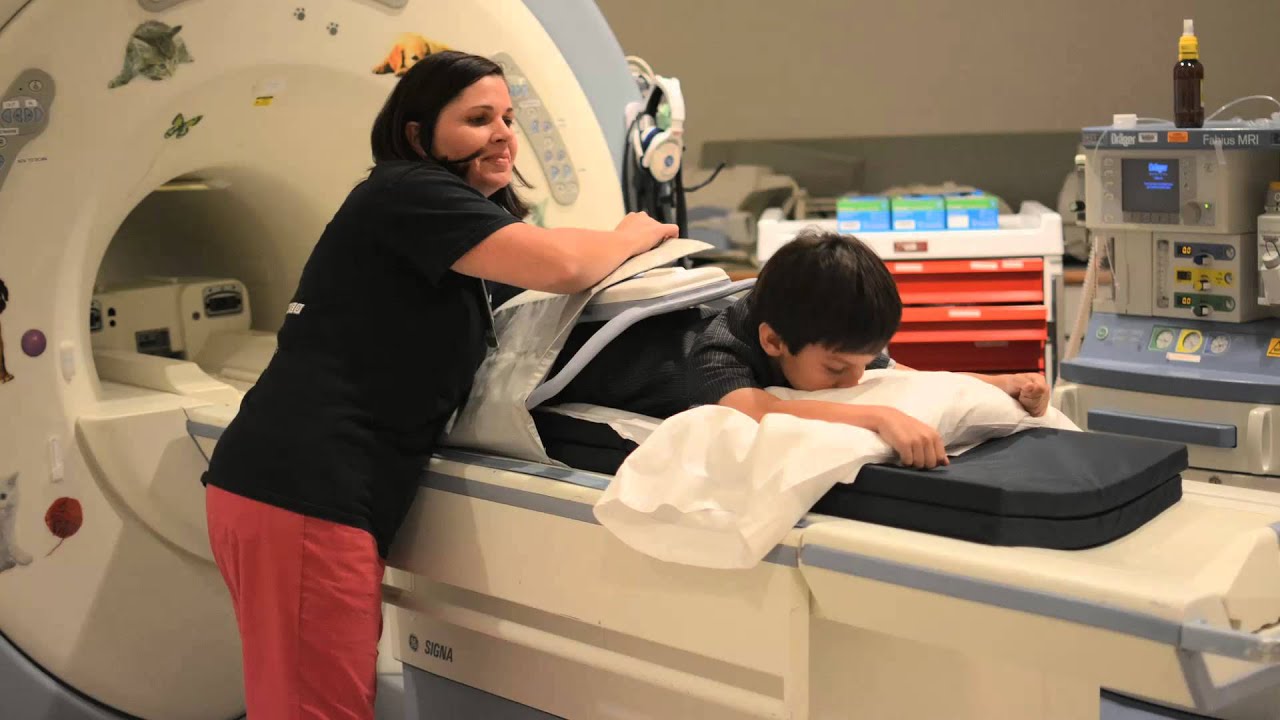
Young children may struggle to remain motionless during the CT scan. If this is the case, the youngster may be given sleeping medication first. This is carried out at the Pediatric Care Unit. If this is done initially, a nurse will also be present for the CT scan. If you are afraid that your small kid may be unable to remain still, speak with your doctor before the CT scan.

After The Test
The weak barium you will consume for this test will not be absorbed by your body and will pass out as a liquid in approximately 2-3 hours. Bowel motions will return to normal after this watery stool has gone. It will help if you drink plenty of water for several hours following the exam.
The CT enterography method has no long-term adverse effects. Because the test does not involve incisions or anesthesia, you will not require recuperation time. You may resume your normal activities once the operation is over. If you are nursing, see your healthcare professional about whether you should postpone breastfeeding for 1 to 2 days. The current state of knowledge is contradictory.
Your test findings will be reviewed by a radiologist and sent to your healthcare practitioner. Your healthcare professional will contact or visit you to discuss the results and future measures.

What to Expect During a CT Scan
Benefits
- CT scanning is noninvasive, painless, and accurate.
- The capacity of CT to scan bone, soft tissue, and blood arteries all at once is a significant benefit.
- CT scanning, unlike traditional x-rays, produces detailed pictures of various kinds of tissue and the lungs, bones, and blood vessels.
- CT scans are quick and easy to do. They can identify internal injuries and bleeding fast enough in an emergency to help save lives.
- CT is a cost-effective imaging method for a variety of clinical situations.
- In comparison to previous small intestinal imaging methods, CT enterography can see the complete thickness of the gut wall and assess adjacent soft tissues. Other tests, some invasive, can only scan the inner lining of the small intestine.
- CT enterography has been proved to diagnose and rule out specific conditions/diseases, which may assist you in deciding on your future medical treatment.
- CT enterography might reduce the requirement for video capsule endoscopy (VCE) and its associated risks.
- Other organs in the abdomen may be visualized with CT enterography.
- CT scans are less sensitive to patient movement than MRI scans.
- Unlike an MRI, having a medical device implanted will not preclude you from getting a CT scan.
- After a CT scan, no radiation is left in the patient's body.
- CT scanning x-rays should have no acute adverse effects.
Risks
- There is always a minor risk of cancer from excessive radiation exposure. The advantage of a precise diagnosis, on the other hand, considerably surpasses the danger of CT scanning.
- If a woman suspects she is pregnant, she should inform her doctor and the x-ray or CT technician. More information on pregnancy with x-rays may be found on Safety in X-ray, Interventional Radiology, and Nuclear Medicine Procedures.
- Because of the possible harm to the unborn baby, doctors typically do not suggest CT screening for pregnant women unless medically required.
- According to IV contrast manufacturers, moms should not nurse their kids for 24 to 48 hours after receiving contrast material. However, according to the most current American College of Radiology (ACR) Manual on Contrast Media, studies reveal that the quantity of contrast absorbed by the newborn while nursing is exceedingly minimal. Please see the ACR Manual on Contrast Media and associated references for further information.
- The likelihood of primary allergic response to iodine-containing contrast materials is relatively low, and radiology departments are well-equipped to handle them.
- CT enterography may now be conducted with significantly lower radiation doses due to technological advances.
- Because children are particularly vulnerable to radiation, they should only receive a CT scan if it is necessary for a diagnosis. They should not undergo repeated CT scans unless essential. CT scans for children should always be performed using a low-dose approach.
CT Enterography Versus Conventional Modalities
CT enterography provides several benefits over small bowel follow-up and traditional enteroclysis. It may show extraluminal pathology and a luminal disease; the whole small bowel can be viewed without being hampered by overlapping loops; and multiplanar reconstructions are routinely used, boosting diagnostic accuracy and confidence. Consequently, a single CT enterography may replace the requirement for many radiological examinations, enhancing diagnostic efficiency, patient compliance, and, ultimately, radiation exposure. When measuring small intestine motility and the patency of sinus or fistulatracts, fluoroscopic minor bowel investigations continue to outperform cross-sectional approaches.
CT enterography images are commonly used to increase diagnostic confidence in excluding small bowel and extraluminal disease. CT provides higher spatial and temporal resolution, and single breath-hold exams are quick and easy. CT enterography is also less expensive and more widely accessible than MRI enterography, and patients benefit from shorter examination periods and avoidance of MRI-associated claustrophobia. MRI, on the other hand, has higher contrast resolution and may detect fistulae better than CT. In adults, gadolinium-based intravenous contrast used for MR imaging may be safer than iodinated contrast used for CT.
Capsule endoscopy is the ideal first-line diagnostic for "conventional endoscopy-negative" gastrointestinal bleeding because it affords excellent visualization of the small intestine mucosa. Furthermore, capsule endoscopy may be preferable to radiographic studies to diagnose early Crohn's disease and small intestinal neoplasms. Indeed, when there is a strong suspicion of underlying small bowel mucosal illness, capsule endoscopy plays a crucial supplementary function. However, capsule endoscopy cannot examine different mucosal symptoms or consequences of minor bowel illnesses; for example, small bowel Crohn's disease is widely recognized as a disease of both mucosa and mesentery, with varied involvement of both components. Furthermore, mucosal visualization is typically inadequate during capsule endoscopy, and potentially harmful consequences such as capsule retention or, in rare cases, capsule endoscope aspiration might occur. Radiological examinations are preferable in individuals with suspected small bowel stricture or established Crohn's disease.
People Also Ask
What Organs Does A CT Enterography Show?
CT enterography produces comprehensive pictures of the small intestine and structures inside the belly and pelvis by using specific x-ray equipment and an injection of contrast material following the consumption of fluids.
How Long Does A CT Enterography Take?
The scan will take around 5 minutes, and you may be given an IV injection of non-ionic contrast throughout the procedure. You may be requested to hold your breath, so your breathing action does not muddy the photos.
What Is The Prep For A CT Enterography?
Unless otherwise directed by your doctor, you should not eat or drink anything for three hours before your exam. Please continue to take your prescriptions as usual. You will be advised to come 90 minutes early to drink three bottles of beverage for your treatment.
What Happens During A CT Enterography?
During CT Enterography, images of cross-sections or slices of your abdominal tissues, focused on the small intestine, are obtained. Before the scan, you will consume a special kind of weak barium and get an injection in a vein through an IV to highlight the small intestine even more.
Conclusion
In the diagnostic arsenal of minor intestinal problems, CT enterography is a helpful and complementary addition to MR enterography/enteroclysis and capsule endoscopy. CT enterography will play an essential role in the future due to the widespread availability of multidetector-row CT platforms and examination efficiency with excellent patient tolerance. However, this must be balanced with careful monitoring of the cumulative radiation dose.

Suleman Shah
Author
Suleman Shah is a researcher and freelance writer. As a researcher, he has worked with MNS University of Agriculture, Multan (Pakistan) and Texas A & M University (USA). He regularly writes science articles and blogs for science news website immersse.com and open access publishers OA Publishing London and Scientific Times. He loves to keep himself updated on scientific developments and convert these developments into everyday language to update the readers about the developments in the scientific era. His primary research focus is Plant sciences, and he contributed to this field by publishing his research in scientific journals and presenting his work at many Conferences.
Shah graduated from the University of Agriculture Faisalabad (Pakistan) and started his professional carrier with Jaffer Agro Services and later with the Agriculture Department of the Government of Pakistan. His research interest compelled and attracted him to proceed with his carrier in Plant sciences research. So, he started his Ph.D. in Soil Science at MNS University of Agriculture Multan (Pakistan). Later, he started working as a visiting scholar with Texas A&M University (USA).
Shah’s experience with big Open Excess publishers like Springers, Frontiers, MDPI, etc., testified to his belief in Open Access as a barrier-removing mechanism between researchers and the readers of their research. Shah believes that Open Access is revolutionizing the publication process and benefitting research in all fields.

Han Ju
Reviewer
Hello! I'm Han Ju, the heart behind World Wide Journals. My life is a unique tapestry woven from the threads of news, spirituality, and science, enriched by melodies from my guitar. Raised amidst tales of the ancient and the arcane, I developed a keen eye for the stories that truly matter. Through my work, I seek to bridge the seen with the unseen, marrying the rigor of science with the depth of spirituality.
Each article at World Wide Journals is a piece of this ongoing quest, blending analysis with personal reflection. Whether exploring quantum frontiers or strumming chords under the stars, my aim is to inspire and provoke thought, inviting you into a world where every discovery is a note in the grand symphony of existence.
Welcome aboard this journey of insight and exploration, where curiosity leads and music guides.
Latest Articles
Popular Articles