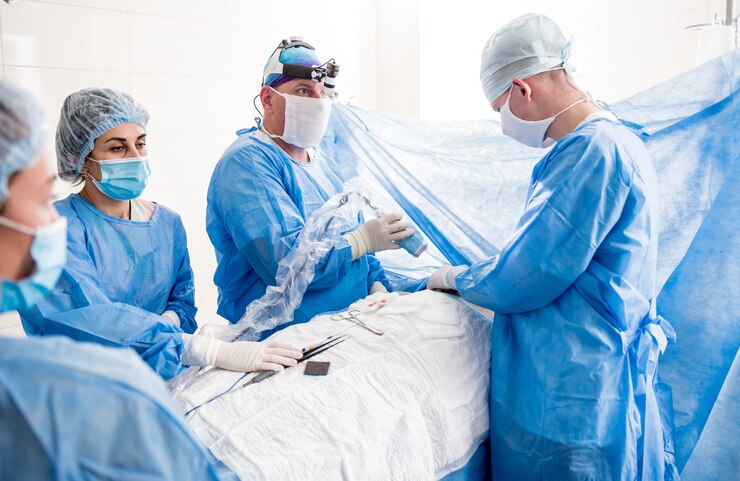
Fluorescence Guided Surgery – Benefits And Future Opportunities
Fluorescence guided surgery is an imaging technology that enables the surgeon to see structures and kinds of tissue that may not be apparent under white light settings during a surgical operation.
Author:Suleman ShahReviewer:Han JuJul 24, 202226 Shares451 Views
Fluorescence guided surgeryis an imaging technologythat enables the surgeon to see structures and kinds of tissue that may not be apparent under white light settings during a surgical operation.
Because of the many potential advantages of fluorescence guided surgery over more traditional clinical imaging techniques, such as higher contrast and sensitivity, less personal use, and ease of instrument operation, research interest in fluorescence-guided surgery continues to grow across a variety of key areas, including fluorescent probe development, surgical system development, and potential clinical applications.
Fluorescence Guided Surgery Principles
In its most basic form, the fluorescence-guided surgery system will consist of a light source and associated filters for excitation of the fluorescence contrast agent, which is often provided before surgery.
The emitted fluorescence signal from the probe is then collected by removing unwanted signals such as excitation light and autofluorescence by first passing the light through appropriate emission filters, followed by collection optics to focus the signal on the detector, where it is transferred to an attached computer for visualization.
Every component in an imaging system impacts the final picture quality in various ways and in varying degrees; therefore, careful thought is necessary depending on the system's use.
While the equipment focus of commercial fluorescence guided surgery systems has hitherto been on fluorescence intensity measurements, there is rising interest in bringing the benefits of fluorescence lifetime to bear, most notably in macroscopic adaptations of fluorescence lifetime imaging microscopy.
Fluorescence Guided Surgery Systems Currently In Use
The FDA has approved roughly 20 fluorescence-guided clinical imaging devices for use in the clinical setting, which usually falls into handheld imaging systems, endoscopic-based systems, and surgical microscopes.
All of them are intensity-based rather than lifetime-based. Fluobeam, which has been used in research applications since 2009, is one of the most advanced FDA-approved systems.
Fluobeam is a portable imaging device with a Class 1 laser beam at 750 nm and emission collection from 800 nm onwards for use with Indocyanine Green in applications such as thyroid and lymphatic surgery.
The da Vinci system is another well-established, endoscopic-based device that incorporates the Firefly fluorescence visualization capabilities and is also intended to function with Indocyanine Green.
In reality, most presently certified systems are intended to function with Indocyanine Green, with excitation occurring at 805 nm and emission captured from 820 onwards.
These systems are often totally enclosed, making access to the optics that need to be updated impossible. Fluobeam can also be used to utilize Methylene Blue fluorescence. Quest Medical Imaging has developed the Quest Spectrum, which can be used for applications utilizing both Methylene Blue fluorescence and Indocyanine Green by incorporating two imaging channels, demonstrating greater versatility than other systems.
The Leica M530 surgical microscope is an even more versatile system, with many accessory modules that can be incorporated into the system for visualization of fluorescence at various wavelengths, including those required for Indocyanine Green and fluorophores with emissions above 450 nm and 510 nm.
Use In Clinical Systems
There is a continual demand for fluorescence-based clinical imaging systems that can work with both authorized fluorophores and those presently under development. Imaging systems may employ a probe's fluorescence intensity, lifespan, wavelength, polarization, and other features in surgery.
Fluorescence lifetime measures independent of probe photobleaching, concentration, and wavelength may reveal more information about a sample than intensity measurements alone.
The research technique of choice is time-correlated single-photon counting, shown in a simplified graphic. Fluorescence lifetime analysis has provided a useful 1 mm phantom tumor margin resolution.
For commercially produced devices, fluorescence intensity remains the preferred measurement. On the other hand, recent advances in single-photon avalanche diode arrays promise to improve the capabilities of fluorescence lifespan measurements considerably.
Components In Fluorescence Guided Surgery
The key instrumentation components for using fluorescence in surgical systems are primarily based on well-established technology and methodologies for fluorescence spectroscopy and microscopy and consist of an excitation source, a fluorescent probe, excitation and emission filters, and the fluorescence detector.
Excitation Sources
Broadband light sources such as xenon lamps, light emitting diodes, and laser diodes are often excitation sources in fluorescence imaging systems. They may not be suitable for use with fluorophores with a tiny Stokes shift, which will need the help of further excitation filtering.
Light emitting diodes are more durable since they do not need a warm-up time and emit across a smaller bandwidth, allowing them to be selected based on the fluorophore being worked with.
Ti: sapphire laser technology has dominated time-resolved fluorescence research for many years, but a white-light supercontinuum laser is another viable source that has not been widely used.
Supercontinuum lasers have a wide spectral range from 400 nm to 2 m and very high intensity. They are also much more expensive than light emitting diodes and are not as small, although they may be employed more in future fluorescence guided surgery systems.
Filters And Collection Optics
The selection of excitation and emission filters enables precise wavelength selection and discrimination of undesirable signals from a sample. When using white light sources, excitation filters may be necessary, while emission filters are utilized to confine the fluorescence signal captured to the spectral region of interest.
Another element to consider when selecting emission filters is the Stokes shift of the fluorophore to be used with the system.
The fluorescence signal gathered from the system is focused onto the detector using collection optics. The optics used will most likely be determined by the desired field of vision, which may be modified by the size of the fiber bundle or light guide.
The kind of detector utilized will also impact the optics selection since the sensor will determine the amount of focus needed.
Fluorescence Detectors
Detectors provide the advantages of detecting photons at the single photon level. A detector's quantum efficiency and intrinsic gain often determine its ability to detect single photons. If a detector was 100 percent efficient, it could create one electron for every photon observed.
Photomultiplier tubes were the first devices to detect single photons. In most cases, photomultipliers use the photoelectric effect, in which light photons enter the input window and are absorbed by a photocathode.
This emission phenomenon is reproduced numerous times on each detector's dynodes. Microchannel plate photomultipliers (MCP-PMs) use photocathodes instead of the photomultiplier tubes chain of dynodes. Because electrons in a microchannel plate are considerably more confined in what route they may take than in dynode photomultiplier tubes, there is less fluctuation in transit time.
Charge-coupled devices (CCDs) are a better option for photomultiplier tubes, especially for clinical applications. Electron multiplying-CCD cameras allow on-chip amplification of the signal and bypass readout noise to retain high sensitivity at fast speeds. The primary benefit of electron multiplying-CCDs over photomultiplier tubes is their ability to provide live imaging.
A single-photon avalanche diode is a photodiode with a reverse biased p-n junction at a voltage greater than its breakdown voltage. When a single photon hits the active device, it generates an electron-hole pair, which starts a self-sustaining avalanche of carriers. The QuantiCam, built by Robert Henderson's team in Edinburgh, is an example of a single-photon avalanche diode array for fluorescence lifetime imaging microscopy.
Fluorescence Guided Surgery Applications
Fluorescence guided surgery offers a broad range of therapeutic uses and generates much research interest for future applications. Some of the essential fluorescence guided surgery applications and potential future uses are described below.
- Mapping of Sentinel Lymph Nodes:Oncology is one of the most popular fields of surgery where fluorescence has been effectively used. One of the most widely studied uses of fluorescence-based surgical proceduresis the ability to detect tumor drainage locations versus lymph nodes reliably. A radioactive tracer and a blue fluorescent dye are typically used to identify and see them. Blue dyes may induce allergy, as well as tattooing and skin discoloration. Methylene Blue alone may obtain identification rates equivalent to the combination of dye and tracer. Alternative dyes, such as Indocyanine Green and Methylene Blue, are also being studied in areas with the greatest nodal detection rates in breast cancer patients.
- Identification Tumour:During cancer surgery, removing as much malignant tissue as possible is critical while avoiding excessive injury to adjacent good tissue. Slide-based histology is the current gold standard for evaluating excised tissue. However, it is conducted post-operatively and is frequently a time-consuming and labor-intensive procedure. Before new technologies may be considered for ordinary clinical practice, they must equal the diagnostic accuracy of postoperative histology. Fluorescence-based approaches for intraoperative margin measurement have gained popularity in recent years. Indocyanine Green is a fluorescent dye extensively used in intraoperative fluorescence imaging investigations. The discovery of novel dyes, as well as the improvement of existing dyes, is also of importance for determining tumor margins.
- Use In Angiography:Angiography is a blood vessel imaging method most typically used to monitor the vessels surrounding the heart, brain, lungs, and kidneys. Egas Moniz demonstrated cerebral angiography for the first time in the 1920s, ushering in the method. It may be used to identify numerous eye disorders such as macular degeneration, diabetic retinopathy, and eye cancer, in addition to monitoring blood circulation.
- Use In Endoscopy:Endoscopes are long, thin, flexible tubes containing a fiber bundle of about 50,000 optical fibers. In the 1950s, Basil Hirschowitz showed the use of a flexible fiber optic endoscope, which was the first practical prototype of such fiber bundles. While most endoscopic techniques employ just white light, fluorescence in conjunction with fiber-based methods may also be used.
- Other Applications:Fluorescence may be utilized to see structures such as nerves, ureters, and those in the bile duct during surgery, reducing needless harm to these body areas. Some examples of fluorescence in these areas include using Indocyanine Green during laparoscopic cholecystectomy to see the cystic duct and reduce intraoperative nerve harm. Fluorescence has also been used to measure vascularisation in other anatomical regions of the body, including the parathyroid glands, employing fluorescence-guided glioblastoma imaging. The topic of robotic-assisted surgery is one area of fluorescence guided surgery research where fluorescence might be tremendously valuable. Some previously stated applications have combined fluorescence with robotic surgery, such as recognizing sentinel lymph nodes during laparoscopy and monitoring tumor margins for diseases such as kidney cancer. Researchers are most interested in applications involving the advancement of cancer surgery, both for robotics-based surgery and in the broader field of fluorescence guided surgery.
People Also Ask
What Is Intraoperative Fluorescence?
Fluorescence-guided surgery is an intraoperative optical imaging technique that gives surgeons real-time guidance for tumor delineation.
What Is The Purpose Of Image Guided Surgery?
Image guided surgery is intended to assist doctors in pinpointing the brain tumor. Doctors must accurately locate and remove the brain tumor for doctors to maintain the patient's brain functioning following surgery.
What Commonly Uses Fluorescence Guided Procedures?
Fluorescence-guided surgery is now employed in various surgical scenarios, including sentinel lymph node mapping, solid tumor diagnosis, lymphography, angiography, and anatomical imaging.
How Does Fluorescence Guided Surgery Work?
Fluorescence guided surgery (FGS) is an imaging technology that enables the surgeon to see structures and kinds of tissue that may not be apparent under white light settings during a surgical operation.
Conclusion
There is still much interest, and promise in fluorescence-based surgery approaches for many surgical applications. The capacity to employ fluorescence to detect information at the molecular level is an obvious benefit for incorporating it into ordinary clinical practice.
Fluorescence may be best employed in the surgical setting as a supplement to current surgical procedures rather than completely replacing them. Bridging the gap between these two sectors will also be critical to advancing fluorescence-guided surgery.

Suleman Shah
Author
Suleman Shah is a researcher and freelance writer. As a researcher, he has worked with MNS University of Agriculture, Multan (Pakistan) and Texas A & M University (USA). He regularly writes science articles and blogs for science news website immersse.com and open access publishers OA Publishing London and Scientific Times. He loves to keep himself updated on scientific developments and convert these developments into everyday language to update the readers about the developments in the scientific era. His primary research focus is Plant sciences, and he contributed to this field by publishing his research in scientific journals and presenting his work at many Conferences.
Shah graduated from the University of Agriculture Faisalabad (Pakistan) and started his professional carrier with Jaffer Agro Services and later with the Agriculture Department of the Government of Pakistan. His research interest compelled and attracted him to proceed with his carrier in Plant sciences research. So, he started his Ph.D. in Soil Science at MNS University of Agriculture Multan (Pakistan). Later, he started working as a visiting scholar with Texas A&M University (USA).
Shah’s experience with big Open Excess publishers like Springers, Frontiers, MDPI, etc., testified to his belief in Open Access as a barrier-removing mechanism between researchers and the readers of their research. Shah believes that Open Access is revolutionizing the publication process and benefitting research in all fields.

Han Ju
Reviewer
Hello! I'm Han Ju, the heart behind World Wide Journals. My life is a unique tapestry woven from the threads of news, spirituality, and science, enriched by melodies from my guitar. Raised amidst tales of the ancient and the arcane, I developed a keen eye for the stories that truly matter. Through my work, I seek to bridge the seen with the unseen, marrying the rigor of science with the depth of spirituality.
Each article at World Wide Journals is a piece of this ongoing quest, blending analysis with personal reflection. Whether exploring quantum frontiers or strumming chords under the stars, my aim is to inspire and provoke thought, inviting you into a world where every discovery is a note in the grand symphony of existence.
Welcome aboard this journey of insight and exploration, where curiosity leads and music guides.
Latest Articles
Popular Articles