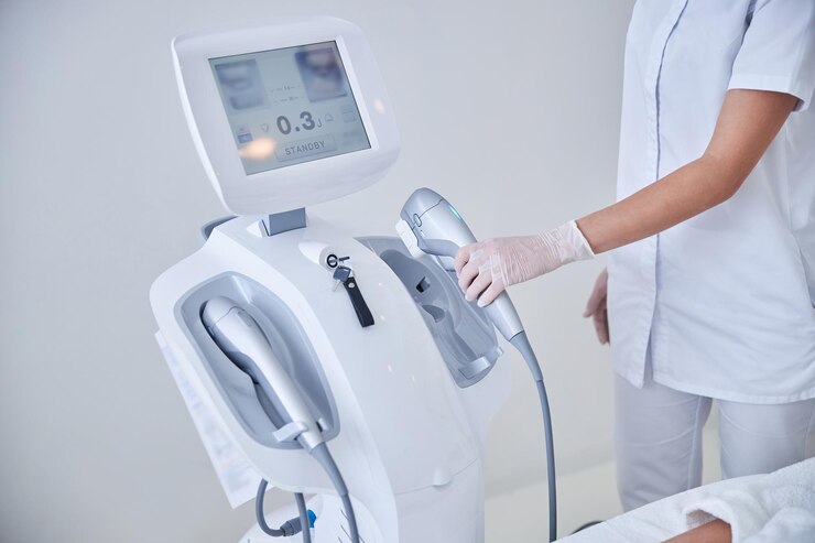
High Intensity Therapeutic Ultrasound – Its Uses And Characterization Techniques
High-intensity therapeutic ultrasound is gaining favor as a promising therapy option for cancers of the uterus, liver, and prostate, among others.
Author:Suleman ShahReviewer:Han JuJul 20, 202289 Shares1.8K Views

High-intensity therapeutic ultrasoundis a non-ionizing, minimally intrusive medical treatment used to treat cancer.
The accurate characterization of high-intensity therapeutic ultrasound fields is critical for patient safety and clinical efficiency. High-intensity therapeutic ultrasound is gaining favor as a promising therapyoption for cancers of the uterus, liver, and prostate, among others.
Because of the rapid growth of high-intensity therapeutic ultrasound applications, there is an urgent demand for accurate measurements of fundamental acoustic parameters and field characteristics. The dependability of high-intensity therapeutic ultrasound field characterization is critical since quality control assurance for the modality is still difficult.
The severely non-linear distorted waveforms have many harmonic frequency components in high-pressure, high-intensity therapeutic ultrasound fields. Better spatial resolution, broad bandwidth, and durability to tolerate probable cavitation effects in powerful areas are in high demand.
Researchers all across the globe are working to develop a standard for field characterization of high-intensity therapeutic ultrasound systems at or near maximum clinical output power. This article examines the state-of-the-art in ultrasonic field characterization methods, emphasizing the commonalities between them.
However, defined quality assurance standards for high-intensity therapeutic ultrasound have yet to be created, a precondition for the widespread adoption of high-intensity therapeutic ultrasound as a treatment modality.
Measurement Of The Pressure Field
The output of a hydrophone is the convolution of the acoustic pressure waveform and its intrinsic system impulse response. An accurate hydrophone would have a consistent frequency response across a broad bandwidth, making it an excellent point receiver.
It would have a high sensitivity to reduce the influence of measurement noise on the uncertainty of findings, as well as a linear dynamic range large enough to identify the peak pressure amplitudes.
However, meeting all of these somewhat contradictory requirements at the same time is unachievable, and appropriate compromises must be established.
Local high pressures, high-speed jets, and shock waves created by cavitation collapse in high-intensity therapeutic ultrasound fields may damage used piezoceramic or piezoelectric hydrophones.
Heat production from absorption in any underlying structure might pose further issues, such as depolarization of the piezoelectric parts.
Thus, researchers have created many hydrophones to increase the measuring capabilities and durability of the hydrophone against high sound intensities and pressures.
Robust Needle Hydrophone
Needle hydrophones are typically made and composed of a disc-shaped active element held at the tip of a needle-like structure. Piezoelectric ceramic or polyvinylidene fluoride are the most often utilized materials for needle hydrophone-sensitive components.
Although the unpredictability of ceramic frequency response may be mitigated, radial resonances should also be addressed with polyvinylidene fluoride-based sensors.
A projectile-like capsule-type hydrophone has been devised to lessen the disruption of diffracted edge waves. A metallic coating was applied to the small piezoceramic sensing element.
The coating thickness was planned to be between 20 and 70 microns to provide a balance between acoustic responsiveness and 'blast protection. The hydrophone's performance stays steady with compressional and rarefactional pressures up to 30 MPa and a pulse burst of 350 cycles.
Robust Membrane Hydrophones
Membrane hydrophones consist of an acoustically transparent polyvinylidene fluoride membrane stretched across an annular frame of internal diameter, typically approximately 100 mm. The major advantage of membrane hydrophones is the broadband and smooth frequency response, which makes them a preferable device for characterizing pulse pressure waveforms.
One method to protect the electrodes from the erosion effect of cavitation is sandwiching the thin membrane between layers of glycerin, which may result in non-flat frequency response. Physikalisch-Technische Bundesanstalt has also developed a robust spot-poled membrane hydrophone for high-intensity therapeutic ultrasound fields characterization.
This hydrophone is reported to have a relatively flat frequency response of up to 15 MHz and can withstand acoustic pressure up to 100 MPa under pulse excitations.
Different protection systems have been used for the rear side of the hydrophone, such as a polymer film protection layer and polyurethane backing in connection with a ring design.
This hydrophone can characterize high-intensity therapeutic ultrasound fields with peak compressional and rarefactional pressures of 75 MPa and 15 MPa, respectively. It uses highly viscous silicone oil with a high cavitation threshold as a liquid backing material.
The sensitivity can be adjusted to the desired pressure amplitude range of detection by modifying the design of the integrated preamplifier.
Reflective Scatter Hydrophone
The brittle electrode pattern and cavitation-induced erosion of the piezoelectric element are widely accepted causes of hydrophone degradation in hostile ultrasonic environments.
One option is to utilize protective layers to boost the robustness against cavitation inside robust needle and membrane hydrophones.
An approach is to use a reflector to disperse the incoming acoustic waves and then detect the scattered field to avoid exposing the sensitive part to the strong field fully.
Using scattering theory, the hydrophone was invented to detect acoustic pressure in the focus zone.
The scatterer is a fused silica optical fiber with a protective outer polyamide covering that may be recut and replaced when damaged.
According to the manufacturer, the receiver array has an adequate sensitivity of 318 dB (1 mV Pa1) before pre-amplification and a bandwidth of more than 15 MHz.
Fiber-optic Hydrophones
Piezoelectric hydrophones are subject to right refractive interference and have a limited effective sensitivity area when used to measure ultrasonic fields. Concerns about the hydrophone's capacity to endure the corrosive environment found in high-intensity therapeutic ultrasound fields have been raised.
Refractometry and interferometry are two optical methods often employed to detect ultrasound. The alteration in the optical mean free path or the optical wavelength may modify the interference pattern depending on the interferometric setups used.
The fiber-optic hydrophone was used to characterize the shock wave fields of lithotripsy. The displacement of the fiber tip caused by a shock wave might reach several optical wavelengths, resulting in significant phase modulation.
Fiber sensors based on optical resonators are a possible alternative to the two-beam interferometer setup. The creation of micron-scale resonators allows the whole sensing system to be miniaturized. Fabry–Perot hydrophones have an excellent spatial resolution of 10 m and a large bandwidth of over 50 MHz.
A multilayer optical fiber optic sensor has been created with a smooth frequency response across a wide frequency range of 1 MHz to 75 MHz. It also features peak-compressional acoustic pressure linearity of up to 37 MPa and less than 5% derivations.
Spatial Averaging Of Hydrophones
Waveform distortion is caused by spatial averaging effects caused by the effective limiting area required for any pressure sensor device. This becomes more relevant as the harmonic frequency content increases at high acoustic pressures.
Hydrophones' spatiotemporal transfer function is often separated into frequency response and spatial averaging. The 'stiff baffle' model may overstate the adequate size at low kb values.
Receiving hydrophones should have comparable directivity patterns as emitters, according to reciprocity.
These models agree well at larger kB values and for small angles of incidence. The 'rigid piston' model may accord better with the experimental data for small-probe-type hydrophones.
Broadband near-field plane-wave pulses were used to test acoustic receivers' directional modulus and phase responses simultaneously. Ultrasound sources created by lasers may provide an alternate option.
Techniques For Optical Detection In Free Space
The noninvasive and efficient characterization of ultrasonic fields is of interest, and optical-based detection methods may provide a better answer.
The probe light in free space optical detection methods is sent in free play rather than restricted in optical fibers as in fiber optic detection techniques.
Refractometry-based approaches are categorized into intensity demodulation and light diffraction tomography, depending on the transduction mechanism used to detect optical changes caused by ultrasound.
- Optical Interferometry:Because the optical light in the two-beam and Fabry–Perot hydrophones is directed via optical fibers, the interferometer may be downsized or built as a robust and adaptable system. A tiny plastic reflecting pellicle is placed into the ultrasonic field to track the motion of acoustic waves. The acoustic pressure achieved in the setup is on the order of several MPa, and the pellicle diameter is roughly 80 mm. Michelson homodyne interferometry is actively path-stabilized at the quadrature working points to produce the best signal. An arc-tangent technique may be used to demodulate the carrier signal. The carrier's quantization noise leads to a less dependable outcome for digital demodulation. Photodetector circuits with larger bandwidths may be necessary to catch the high-frequency carrier signal.
- Optical Refraction Technique:Because the speed of sound is significantly slower than the speed of light, the local acoustic pressure maximum and minimum seem to remain stationary for light as it propagates through the sound field. Ultrasound has the properties of an optical phase grating. Acoustic-optical interactions include Raman-Nath diffraction, Bragg diffraction, and light deflection. The refractive index fluctuates drastically in the focus zone of high-intensity therapeutic ultrasound fields, deflecting the light beam. The maximum pressure measured using light deflection is 16.7 MPa, possibly detecting considerably greater acoustic pressure. A complete analytical model for extremely nonlinear fields is currently in the works.
- Schlieren, Shadowgraphy, And Chase contrast:Because the speed of sound is significantly slower than the speed of light, the local acoustic pressure maximum and minimum seem to remain stationary for light as it propagates through the sound field. Ultrasound has the properties of an optical phase grating. Acoustic-optical interactions include Raman-Nath diffraction, Bragg diffraction, and light deflection. The refractive index fluctuates drastically in the focus zone of high-intensity therapeutic ultrasound fields, deflecting the light beam. The maximum pressure measured using light deflection is 16.7 MPa, possibly detecting considerably greater acoustic pressure. A complete analytical model for extremely nonlinear fields is currently in the works.
- Light Tomography:Light diffraction tomography and light refractive tomography are the two types of light tomography used to detect ultrasound fields. Light diffraction tomography is based on Raman-Nath diffraction. Laser beams with beam widths comparable to ultrasound wavelengths pass through the ultrasound field and are diffracted by an ultrasonic phase grating. Right refractive tomography detects the ultrasonic field with a narrow laser beam and has the advantage of a simplified optical path adjustment. Instead of the Klein-Cook parameter or the severe requirement for Bragg incidence encountered in light diffraction tomography, the laser beam width determines light refractive tomography accuracy.
Infrared Thermography Technique
The localized heating impact of high-intensity therapeutic ultrasound fields in a plane of interest may be captured via thermal imaging. As a result, it is a possible substitute for hydrophone scanning. Thermal imaging employs an infrared camera to measure the temperature distribution caused by absorbing ultrasonic waves on tissue-mimicking material.
The goal of temperature monitoring is to determine the geographical distribution of time-averaged intensity in the high-intensity therapeutic ultrasound field. The main challenge of this technologyis the convective heat transfer between the air and the absorbent substance.
Fast temperature fluctuations cause rapid convection of thermal energy. The diffusion over the tiny acoustic beams makes quantitative analysis difficult.
Later, the model was tested experimentally and compared to hydrophone measurement findings, with the agreement dependent on the transducer's focal gain.
The top acoustic pressure limit was set at 3.8 MPa, which was believed to be within the linear acoustic range. Other quantitative approaches for infrared thermography include using a hydrophone to link temperature increase with pressure distributions obtained from a hydrophone.
The thermal distribution over the area of interest may be acquired by moving the infrared camera and tissue-mimicking material along the acoustic axis using an automated stage.
Measurement And Modeling-Based Combined Method
Direct measurement of clinical high-intensity therapeutic ultrasound fields places stringent constraints on the resilience of hydrophones in the presence of strong fields. Researchers used Khokhlov-Zabolotskaya-Kuznetsov modeling to characterize the fields of single-element focusing transducers.
Planar hydrophone scanning is utilized in a more advanced way to describe random arrays of components. This approach suits a broader range of high-intensity therapeutic ultrasound source geometries and pressure distributions.
These optical technologies can potentially measure the near-field hologram or 3D pressure distribution faster than scanning a hydrophone, and the notion of combining measurement and modeling can then be used.
Optical systems based on acousto-optical interactions may suffer from weak interactions and have a dynamic range confined to comparably low acoustic pressure.
Meanwhile, evanescent waves may be ignored in Rayleigh integrals without losing essential information, and the angular spectrum is computationally more efficient.
People Also Asked
What Is Therapeutic Ultrasound Good For?
Your physical therapist may utilize therapeutic ultrasound to deep heat soft tissue and enhance blood flow to certain tissues. In theory, this might improve healing and reduce discomfort.
Is High-Intensity Focused Ultrasound Safe?
High-intensity focused ultrasound has a very good safety profile. The risk of skin burn is the most serious. Cooling devices and treatments are utilized to protect the skin throughout the treatment procedure to reduce this danger.
What Is Considered High-Intensity Ultrasound?
High-intensity focused ultrasound is a non-invasive therapy that employs concentrated ultrasound waves to thermally ablate a piece of tissue, destroying it with tremendous heat.
What Is High-Intensity Focused Ultrasound Used For?
High-intensity focused ultrasound is a novel, non-invasive therapy for prostate cancer and pain caused by cancer that has spread to the bones. The ultrasonic transducer used in high-intensity focused ultrasound is similar to those used in diagnostic imaging, but it generates significantly stronger sound waves.
Conclusion
Over the last decade, the development of novel sensor technology, the development and standardization of complex-valued frequency response calibration of hydrophones and deconvolution procedures, as well as the associated uncertainty determination, may have all contributed to more reliable and efficient high-intensity therapeutic ultrasound field characterization, thereby promoting the modality's clinical applicability.

Suleman Shah
Author
Suleman Shah is a researcher and freelance writer. As a researcher, he has worked with MNS University of Agriculture, Multan (Pakistan) and Texas A & M University (USA). He regularly writes science articles and blogs for science news website immersse.com and open access publishers OA Publishing London and Scientific Times. He loves to keep himself updated on scientific developments and convert these developments into everyday language to update the readers about the developments in the scientific era. His primary research focus is Plant sciences, and he contributed to this field by publishing his research in scientific journals and presenting his work at many Conferences.
Shah graduated from the University of Agriculture Faisalabad (Pakistan) and started his professional carrier with Jaffer Agro Services and later with the Agriculture Department of the Government of Pakistan. His research interest compelled and attracted him to proceed with his carrier in Plant sciences research. So, he started his Ph.D. in Soil Science at MNS University of Agriculture Multan (Pakistan). Later, he started working as a visiting scholar with Texas A&M University (USA).
Shah’s experience with big Open Excess publishers like Springers, Frontiers, MDPI, etc., testified to his belief in Open Access as a barrier-removing mechanism between researchers and the readers of their research. Shah believes that Open Access is revolutionizing the publication process and benefitting research in all fields.

Han Ju
Reviewer
Hello! I'm Han Ju, the heart behind World Wide Journals. My life is a unique tapestry woven from the threads of news, spirituality, and science, enriched by melodies from my guitar. Raised amidst tales of the ancient and the arcane, I developed a keen eye for the stories that truly matter. Through my work, I seek to bridge the seen with the unseen, marrying the rigor of science with the depth of spirituality.
Each article at World Wide Journals is a piece of this ongoing quest, blending analysis with personal reflection. Whether exploring quantum frontiers or strumming chords under the stars, my aim is to inspire and provoke thought, inviting you into a world where every discovery is a note in the grand symphony of existence.
Welcome aboard this journey of insight and exploration, where curiosity leads and music guides.
Latest Articles
Popular Articles
