The Zygomatic Ligament - Critical Details Associated With The Face
In 1959, the zygomatic ligament was first described by McGregor. He discussed this anatomical structure in relation to his surgical techniques for facial rejuvenation during the Annual Meeting of the California Society of Plastic Surgeons in Yosemite, California.
Author:Suleman ShahReviewer:Han JuJun 20, 20226 Shares276 Views

In 1959, the zygomatic ligamentwas first described by McGregor. He discussed this anatomical structure in relation to his surgical techniquesfor facial rejuvenation during the Annual Meeting of the California Society of Plastic Surgeons in Yosemite, California.
When Mrs. Rosebud Preddy, a medical artist at the Letterman General Hospital in San Francisco, California, prepared a slide presentation for Dr. Mar McGregor, she dubbed the ligament 'McGregor's patch.' Since then, the denomination has endured. 'An area of fibrous attachment between the anterior edge of the parotid fascia and the dermis of the cheek skin,' according to McGregor. The undermining skin was not to be extended beyond this patch as part of his facelift procedure.
The zygomatic ligament has largely been studied by plastic surgeons. Descriptive studies (outcome studies) make up the majority of their publications.
Furnas published the first thorough description of the anatomy of the facial ligaments in 1989. 'A bundle of white firm fibers, 3 mm in width and 0.5 mm in thickness, located 4.5 cm in front of the tragus,' according to the zygomatic ligament's description.
Similar descriptions have been given for the tissues of the breast and the feet. In 1992, Stuzin et al. elaborated on the concepts described by Furnas.
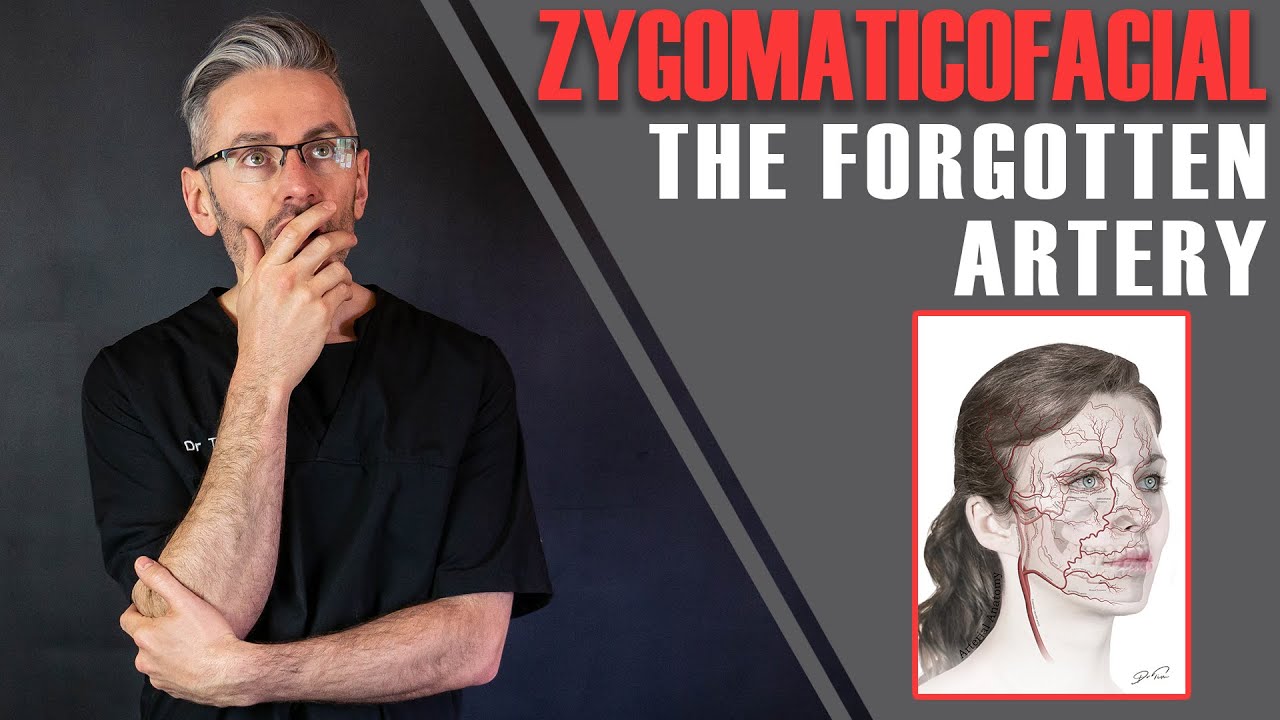
The Forgotten Artery: Why You SHOULD Know About Zygomaticofacial [Aesthetics Mastery Show]
Descriptive Anatomy
The zygomatic, masseteric (or parotidomasseteric), mandibular, and platysma auricular ligaments are the four main ligaments in the face. The preauricular parotid, SMAS malar, and anterior platysma cutaneous ligaments are some of the other minor structures in the face. These ligaments, which run from deep within the dermis to the overlying layers of the skin through the retinacula cutis, fix facial structures and serve as anchoring units for the face, supporting the soft tissue of the face over the underlying bony structures. The retinacula cutis is a connective tissue made up of numerous small fibrous strands that run through the superficial fascia, connecting the deep surface of the dermis to the underlying deep fascia and determining the skin's mobility over deep structures.
According to Stuzin et al., the zygomatic ligament is not one single structure. The main component of these fibrous tissues that begin laterally where the zygomatic arch joins the body of the zygoma is referred to as the 'McGregor patch' in the original description. This main structure passes through the malar fat pad and into the malar skin above. Near the zygomaticomaxillary suture, an isolated structure emerges from the most medial part of the zygoma. These septa connect to form a framework that prevents the face from being shaved. For the human face, this framework acts as a "retaining system."
Superficial Anatomy
According to Furnas, the zygomatic ligaments are located 4.5 cm ahead of the tragus. Rossell-Perry et al. found the zygomatic ligament to be 4.3 cm to 5.5 cm in front of the tragus (average: 5.0 +/-0.336) in a recent study. Ozdemir et al. found the location of the zygomatic ligament to be 4.2 cm to 4.8 cm in front of the tragus in men and 3.9 cm to 4.5 cm in women.
Younger people are less aware of this, while the elderly are more aware. According to Furnas, the proximal insertion is typically unrelated to age and remains unchanged throughout the aging process. The location of the zygomatic ligaments was found to be slightly affected by age, according to Raksin et al. The zygomatic ligaments' location varies with age (distal insertion only), sex, race, and population. For these variables, no research has been done on the anatomic variations of the zygomatic ligament.
Relations
The zygomatic ligaments have the following important relationships: 1) above, the prezygomatic space and the zygomaticofacial nerve, a branch of the maxillary division of the trigeminal nerve; 2) medially, the buccal branch of the facial nerveto the orbicularis oculi muscle, the infraorbital nerve, and the malar fat pad; and 3) below, the upper zygomatic branch of the facial nerve, zygomatic muscles.
Function
The importance of the zygomatic ligament stems from its ability to suspend malar soft tissue over the zygomatic eminence. The effectiveness of the ligament is determined by the width of its skin attachments, and if the attachments are too small, the ligament will stretch over time. The lack of ligament support, which is one of the main causes of an aging face, causes the malar fat pad to move downward. The thin anterior wall of the maxilla and the maxillary sinus are protected from trauma by the malar fat pad.
These septa and ligaments, according to Rohrich et al., help to keep the blood supply to the skin stable. The septal membranes are found between anatomical compartments in vascular zones. Perforators from the transverse facial artery and the angular artery can be found in the medial face. The zygomatic ligaments may also help to prevent ectropion of the lower eyelid. Lower eyelid ectropion can result from defects of the midface that lack the zygomatic ligament and are repaired with flaps.
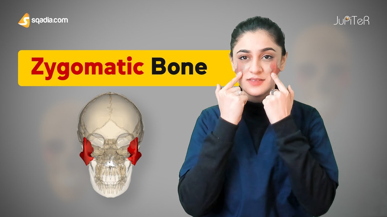
Zygomatic Bone | Cranial Osteology | Anatomy Lecture for Medical Students | V-Learning™
Innervation
The zygomatic ligaments receive sensation from branches of the zygomaticofacial nerve and the infraorbital nerve.
Blood Supply
The zygomatic ligaments are supplied with blood via branches of the transverse facial artery, angular artery, and infraorbital artery. The venous plexus, which includes the transverse facial vein and the facial (angular) vein, is where venous drainage takes place.
Clinical Applications
Cosmetic surgery is the most common use for these anatomical structures. Face rejuvenation procedures include surgical correction of the retaining ligaments, plication of the SMAS, and repositioning of the soft tissues of the face that have descended with gravity. The restoration of the malar soft tissue, as well as repositioning the tissue to its previous position, should be used to treat facial aging of the nasolabial folds. Aufricht, Stuzin, Muzzafar et al., Mendelson et al., Ozdemir et al., and other plastic surgeons have reported improvements in the aging of the face based on ligamentary suspension of the soft tissues.
The surgical anatomyof the facial retaining ligaments and its significance in facelift surgery have been described by these authors. Aston described fingertip dissection of the malar fat pad through the prezygomatic space as a technique for midface elevation and suspension. Rossell-Perry et al. recently published a technique for improving aging of the nasolabial folds and neck using SMAS plication to facial ligaments.

People Also Ask
What Do Facial Ligaments Do?
These ligaments serve as anchor points, securing the skin and superficial fascia (SMAS) to the underlying deep fascia and facial skeleton at specific anatomic locations.
Are There Ligaments In Your Face?
The retaining ligaments of the face are thick, deep fibrous attachments that originate in the periosteum or deep facial fascia and travel perpendicularly through facial layers to the dermis.
What Are The True Retaining Ligaments Of The Face?
Currently, the orbital, zygomatic, and mandibular ligaments (LaTrenta) are considered the true retaining ligaments of the face. Between the superolateral orbital rim and the superficial temporal fascia (Knize), the orbital ligament is located.
What Is Zygomaticus Major?
The zygomaticus major muscle is perhaps the most visible of all the facial muscles. It controls the way we smile because it sits between the corners of our lips and the upper part of our cheeks. The zygomatic bone, also known as the cheekbone, sits on top of the muscle.
Conclusion
The zygomatic ligament is a group of structures that work together. The main component of this series of fibrous tissues that begin laterally where the zygomatic arch joins the body of the zygoma is referred to as the 'McGregor patch' in the original description. The prezygomatic space, zygomatic branches of the facial nerve, and zygomaticofacial nerve are the three most important relationships of this structure. Cosmetic surgeries (facelifts) are the most common use for these anatomical structures. More research is needed to better understand the relationship between the zygomatic ligament and factors like age, sex, race, and other factors.

Suleman Shah
Author
Suleman Shah is a researcher and freelance writer. As a researcher, he has worked with MNS University of Agriculture, Multan (Pakistan) and Texas A & M University (USA). He regularly writes science articles and blogs for science news website immersse.com and open access publishers OA Publishing London and Scientific Times. He loves to keep himself updated on scientific developments and convert these developments into everyday language to update the readers about the developments in the scientific era. His primary research focus is Plant sciences, and he contributed to this field by publishing his research in scientific journals and presenting his work at many Conferences.
Shah graduated from the University of Agriculture Faisalabad (Pakistan) and started his professional carrier with Jaffer Agro Services and later with the Agriculture Department of the Government of Pakistan. His research interest compelled and attracted him to proceed with his carrier in Plant sciences research. So, he started his Ph.D. in Soil Science at MNS University of Agriculture Multan (Pakistan). Later, he started working as a visiting scholar with Texas A&M University (USA).
Shah’s experience with big Open Excess publishers like Springers, Frontiers, MDPI, etc., testified to his belief in Open Access as a barrier-removing mechanism between researchers and the readers of their research. Shah believes that Open Access is revolutionizing the publication process and benefitting research in all fields.

Han Ju
Reviewer
Hello! I'm Han Ju, the heart behind World Wide Journals. My life is a unique tapestry woven from the threads of news, spirituality, and science, enriched by melodies from my guitar. Raised amidst tales of the ancient and the arcane, I developed a keen eye for the stories that truly matter. Through my work, I seek to bridge the seen with the unseen, marrying the rigor of science with the depth of spirituality.
Each article at World Wide Journals is a piece of this ongoing quest, blending analysis with personal reflection. Whether exploring quantum frontiers or strumming chords under the stars, my aim is to inspire and provoke thought, inviting you into a world where every discovery is a note in the grand symphony of existence.
Welcome aboard this journey of insight and exploration, where curiosity leads and music guides.
Latest Articles
Popular Articles